


Call us for any question
(720) 893-5000Clinic Contact
(720) 772-8040805 E 144th Ave suite 200
Thornton, CO 80023

An ultrasound uses high-frequency sound waves to capture pictures of the inside of the body. It allows your doctor to see real-time images of the movement of the body’s internal organs, vessels, and tissues without making an incision.
A diagnostic ultrasound is used to provide visualization of the structures within your body to provide valuable information for diagnosing and treating a variety of diseases and conditions. It is a useful way of examining many of the body’s internal organs, including:
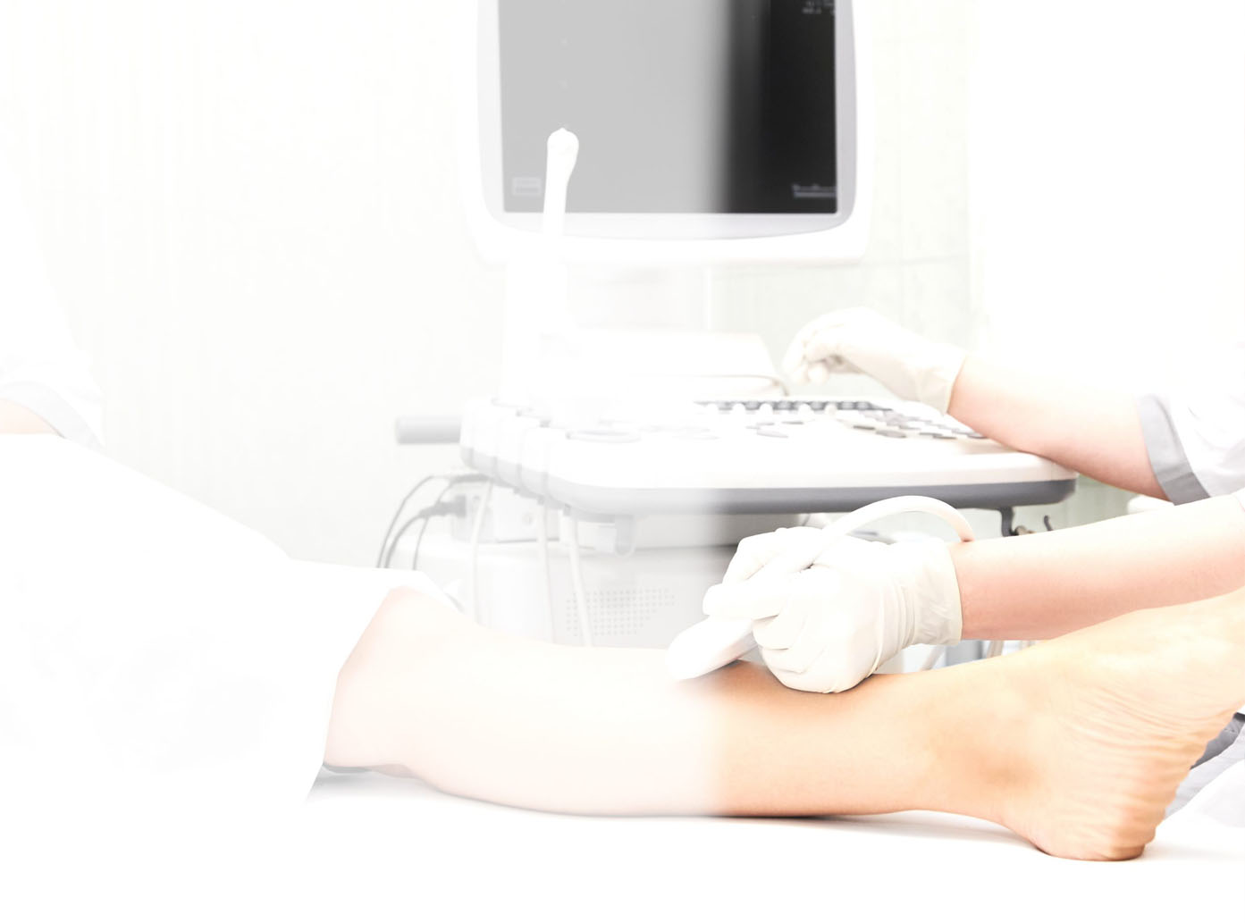
You will change into a hospital gown and lie down on a table. An ultrasound technician, called a sonographer, will apply a gel to the skin over the area being examined. This gel is used to prevent or reduce air pockets, as sound waves have a difficult time traveling through the air.
The transducer, a small, handheld instrument, is pressed against the area being studied and captures the images. Sound waves echo as they pass through the body and then are reflected back into a computer, creating images.
After the ultrasound, the sonographer will clean the gel off your skin. The entire procedure typically takes less than 30 minutes, depending on the area being examined.